When assessing dense breast tissue, it is crucial to use AI that is both objective and automated.
The subjectivity inherent in human evaluations can lead to variability and inconsistency, potentially impacting patient outcomes. AI algorithms, on the other hand, provide standardized, reproducible assessments, ensuring that breast density measurements are accurate and reliable.
Limitations of visual assessment
Visual assessment of breast density is fraught with challenges due to significant reader variability and inconsistency.
Radiologists often rely on subjective judgment when evaluating mammograms, leading to discrepancies in density categorization. This inconsistency can result in varied interpretations for the same patient, affecting clinical decisions and follow-up recommendations.
Variability among radiologists
Radiologist experience level further compounds the variability in density assessment.
Quick fact:
- One study examined 83 radiologists who reviewed over 200,000 mammograms and showed substantial variation in density assessments, with some radiologists classifying as few as 6% and others as many as 85% of mammograms as dense.
This variability, even after adjusting for factors like age and BMI, highlights the inherent subjectivity in visual assessments and emphasizes the need for more consistent, objective methods.
Limitations of area-based density assessment
Area-based density assessments, which focus on the proportion of dense tissue visible in a 2D mammographic image, have inherent limitations.
These assessments are binary, often categorizing breasts as either dense or not without accounting for the volume of dense tissue. Such an approach fails to capture the three-dimensional nature of breast tissue, leading to an incomplete understanding of the masking risk dense tissue poses or the risk of developing breast cancer.
Vendor processing and standardization
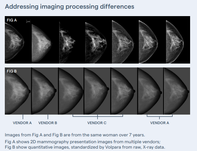
One significant challenge in breast density assessment is the lack of standardization in image processing across different vendors. This processing is applied to maximize contrast to help detect relevant signs of cancer, but it can also distort the visualization of density and lead to inconsistencies in density evaluation across vendors.
Addressing these challenges requires collaboration among vendors, radiologists, and AI developers. Unlike other approaches, Volpara works directly with the raw images that haven’t been altered by processing, ensuring more consistent and accurate breast density assessments and supporting standardized protocols for more reliable results.

How explainable AI can bridge the gap
To overcome these limitations, there is a growing need for explainable and generalizable AI algorithms in breast density assessment. Such algorithms can provide consistent, objective, and reproducible results, reducing the variability seen with human readers.
AI can understand the complexities of breast tissue composition beyond simple visual assessment and offer a more nuanced evaluation of density.
Quantitative measures and unprocessed image data in AI
Volpara leverages quantitative measures and unprocessed image data to assess breast density more accurately. The unprocessed data retains the true X-ray attenuation to pixel brightness relationship data that AI can quantify the amount of dense tissue and its distribution by analyzing pixel-by-pixel variations and patterns within mammographic images.
This method provides a volumetric assessment of density, capturing the true extent of fibroglandular tissue and offering a clearer picture of potential masking risk and lifetime risk of breast cancer.
Why volumetric density assessment?
A volumetric density assessment measures the actual volume of fibroglandular tissue in the breast using quantitative imaging data rather than just the 2D area seen on mammograms.
Volumetric density assessments provide a more comprehensive and accurate picture of breast density, accounting for the depth and distribution of dense tissue. Unlike the broad four-category BI-RADS system, volumetric assessments are key to delivering personalized results, offering better risk prediction for developing breast cancer.
As a result, they offer better risk prediction for breast cancer by more precisely identifying the masking risk of dense tissue and its potential to hide tumors, leading to more personalized and effective screening strategies.
Volpara’s TruDensity® Algorithm
Volpara’s TruDensity algorithm exemplifies the advancement in breast density assessment technology.
Developed using a combination of X-ray physics and machine learning, it generates an accurate and objective volumetric measure of breast composition. This algorithm considers various image parameters and provides consistent density readings regardless of the radiologist’s experience level.
TruDensity’s volumetric approach offers a comprehensive understanding of breast density—improving risk prediction and aiding in personalized screening strategies.
The Volpara Health suite of services offers numerous ways to standardize measurement and assess breast density with precision. Contact us today to consult with one of our breast density experts.
#WorldDenseBreastDay

