First, everyone at Volpara would like to take this opportunity to thank our customers and all healthcare workers and first responders for their extraordinary dedication during the COVID 19 pandemic. We are committed to doing whatever we can to support your efforts in serving our local communities. Our teams across the globe are working remotely to offer virtual training and support, knowing that together we will navigate the unprecedented global challenge before us.
Volpara recently provided an educational grant to Mammography Educators to host a virtual CEU course. Louise Miller, R.T.(R)(M)(ARRT), CRT, FSBI, FNCBC, Director of Education and cofounder of Mammography Educators, presented “What Now? Strategies for Maintaining Image Quality & Patient Care in the Midst of Chaos.”
“This is great time for breast imagers look at their service with new eyes—to look for opportunities to improve image quality going forward,” said Louise. “Focus on what you can do now: utilize resources and reboot ideas for improving efficiency, proficiency, and reproducibility, which improve patient care.”
Whether your breast center has suspended all screening temporarily or is running minimal hours, Louise offered strategies for how to leverage that time now and prepare for increased patient volumes when centers resume regular screening exams.
Louise added: “Improving image quality is one of the most important aspects of patient care. We know that image quality directly affects the ability to detect early cancers; it affects treatment; and most importantly, it affects survival rates.”
So, how can you work on image quality when you’re not working on images? Volpara has committed to making mammography quality and performance data from Volpara®Enterprise™ software remotely accessible to breast centers at any time. This includes access to all training resources, including the positioning video series embedded in the software, to analyze performance and focus training on growth areas.
In the session, Louise offered three key opportunities for improvement during the screening slowdown:
- Image quality analysis. The FDA’s EQUIP initiative provides a means for facilities to focus on real improvement in key areas that impact clinical image quality; namely, patient positioning and compression. Analyze past EQUIP data to discover clues to areas of improvement areas; automated image quality evaluation will help expedite the process.
- Machine calibration. Compare pressure and dose information for all machines. Estimations provided by mammography manufacturers vary according to the algorithm and how they handle breast densities. Work with your vendors to address any calibration issues.
- Workflow analysis. Review this information now to have it available for when patients return. Are all your rooms being utilized at full capacity? Analyze time spent in the room and the number of patients imaged by each unit and by each technologist. Objective data provides the ability to adjust training or staffing to address any issues.
Prep for higher volumes
Louise closed out the session by focusing on how to prepare for higher volumes when screening resumes. Tips included reconfiguring your waiting room to address physical distancing concerns; completing technology and system upgrades; and creative staffing approaches to extend hours (adding evening and/or weekends) to reduce frustration and anxiety as you address backlog.
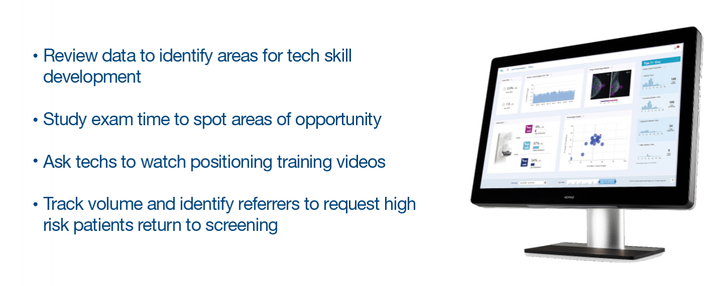
One example of a center that has worked to prepare during this time comes from Katie Kaminski, MRT (R), of Mayfair Diagnostics in Calgary, Canada. Katie is developing a plan for how to use VolparaEnterprise data and analytics tools to track utilization, inform training, and prioritize patients with high breast density. “For example, we will watch the performance data closely to see if techs need work in certain areas as they return to performing exams and encourage them to use the imbedded training videos to refresh their skills,” said Katie.
From an operations standpoint, Mayfair plans to monitor volume compared to pre-COVID and determine when to scale back extra screening times to cover the backlog. “In addition,” Katie concluded, “we can use referring physician data to track where volumes may be down and reach out to those physicians to encourage them to send their patients for screening again. We can also use the referring info to target practices with high volumes of patients with high breast density and educate them on the value of supplemental imaging with ultrasound.”

Stay tuned for a new blog series: Preparing to Reopen Screening Operations
According to Louise: “Nothing should go back to normal; normal wasn’t working. If we go back to the way things were, we will still have missed the lesson. May we rise up and Do Better.”
